


Coxarthrosis affects the hip joints of middle-aged and older people. The causes of its development are previous injuries, congenital and acquired diseases of an inflammatory or non-inflammatory nature. The main symptoms of coxarthrosis are pain in the hip joint, morning swelling and stiffness of movement. In the initial stage of the pathology, treatment is conservative. If it is ineffective against the background of rapid progression of coxarthrosis or its late detection, surgical intervention, usually endoprostheses, is indicated.
Description of the pathology.
Coxarthrosis (osteoarthrosis, deforming osteoarthritis) is a degenerative-dystrophic pathology of the hip joint. At the initial stage of development, the structure of the synovial fluid changes. It becomes slimy, thick and therefore loses its ability to nourish the hyaline cartilage. Due to dehydration, its surface dries out and becomes covered with multiple radial cracks. In this condition, the hyaline cartilage does not soften shocks well when the bones that make up the joint come into contact.
To adapt to the increased pressure that occurs on them, bone structures deform with the formation of growths (osteophytes). The metabolism in the hip joint deteriorates, which negatively affects the muscles and the tendon-ligamentous apparatus of the joint.
Degrees
Each stage is characterized by its own symptoms, the severity of which depends on the degree of narrowing of the joint space and the number of bone growths formed.
| Coxarthrosis severity | Characteristic symptoms and radiological signs. |
|---|---|
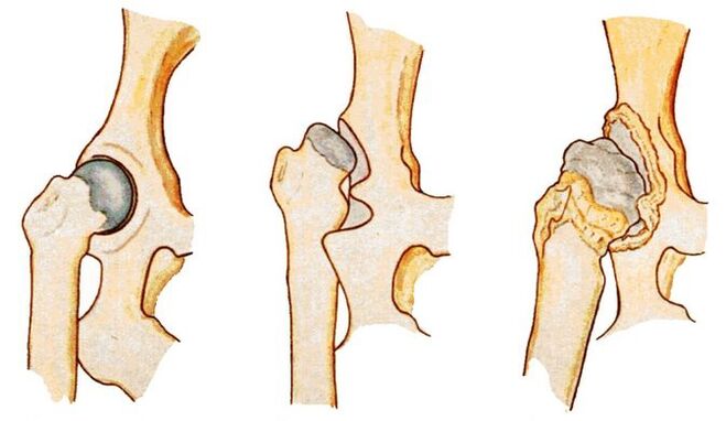
| First | The joint space narrows unevenly and unique osteophytes have formed around the acetabulum. Mild discomfort occurs, but most often the disease does not manifest itself clinically. |
| Second | The joint space is reduced almost 2 times, the head of the femur is displaced, deformed, enlarged, and bone growths are found even outside the cartilaginous lip. Hip pain becomes constant and is accompanied by a significant limitation of mobility. |
| Third | Complete or partial fusion of the joint space, multiple bone growths, expansion of the femoral head. The pain occurs day and night and spreads to the thighs and legs. Movement is only possible with the help of a cane or crutches. |
Causes of the disease
Primary coxarthrosis is a destructive-degenerative injury of the hip joint, the causes of which have not been established. This means that no prerequisites for premature destruction of hyaline cartilage were identified. The following pathological conditions can cause secondary coxarthrosis:
- previous injuries: fracture of the femoral neck or pelvic bones, dislocation;
- hip dysplasia;
- aseptic necrosis of the femoral head;
- congenital hip dislocation;
- inflammatory, including infectious joint diseases (rheumatoid, reactive arthritis, gout, tendonitis, bursitis, synovitis).
Prerequisites for the development of coxarthrosis are obesity, increased physical activity, sedentary lifestyle, metabolic disorders, hormonal disorders, kyphosis, scoliosis and flat feet.
Symptoms of the disease
At the initial stage of development, coxarthrosis can only manifest itself with mild pain. They usually appear after intense physical effort or a hard day of work. The person attributes the deterioration of their health to muscular "fatigue" and does not seek medical help. This explains the frequent diagnosis of coxarthrosis at stages 2 or 3, when conservative therapy is ineffective.
Limitation of joint mobility.
The range of motion in the hip joint is reduced due to compensatory growth of bone tissue, damage to the synovial membrane and replacement of areas of the joint capsule with fibrous tissue devoid of functional activity. Mobility may be somewhat limited even with grade 1 coxarthrosis. Difficulties arise when performing rotational movements with the leg.
As the disease progresses, morning stiffness and joint swelling become common. To regain mobility, a person has to warm up for several minutes. By lunchtime, the range of motion is restored, including as a result of the production of hormone-like substances in the body.
Crunch
When walking, flexing and (or) extending the hip joint, clicking, cracking and crunching sounds are clearly heard. The reason for this accompanying sound with each step is the friction of bone surfaces, including osteophytes, against each other. Crackling sounds can also appear in normal health conditions due to the collapse of carbon dioxide bubbles in the joint cavity. Coxarthrosis is indicated by its combination with dull or sharp pain.
Pain
Painful sensations become constant already at stage 2 of coxarthrosis. Its severity decreases slightly after a long rest. The pain intensifies during the next relapse or the development of synovitis (inflammation of the synovial membrane) that often accompanies osteoarthritis. During the remission stage, the discomfort decreases somewhat. But as soon as a person becomes hypothermic or lifts a heavy object, severe pain appears again.
muscle spasm
Increased tension in the skeletal muscles of the thigh occurs with coxarthrosis for several reasons. First, the ligaments weaken. The muscles spasm to hold the head of the femur in the acetabulum. Secondly, increased tone usually accompanies inflammation of the synovial membrane. Thirdly, when osteophytes are displaced, nerve endings are compressed and muscle spasm becomes a compensatory reaction to acute pain.
Limp
In the later stages of the development of coxarthrosis, the patient begins to limp severely. Changes in gait are caused by flexion contractures and deformation of the bone surfaces, which makes it impossible to maintain the straight leg position. The person also limps to reduce the intensity of the pain by transferring body weight to the unaffected limb.
leg shortening
Shortening of the leg by 1 cm or more is typical for grade 3 coxarthrosis. The reasons for the decrease in the length of the lower extremities are severe muscle atrophy, thinning and flattening of the cartilage, narrowing of the joint space and deformation of the femoral head.
Diagnostic methods
The initial diagnosis is made based on the patient's complaints, external examination, medical history and the results of a series of functional tests. Many inflammatory and non-inflammatory pathologies disguise themselves as symptoms of coxarthrosis, which is why instrumental and biochemical studies are performed.
x-ray examination
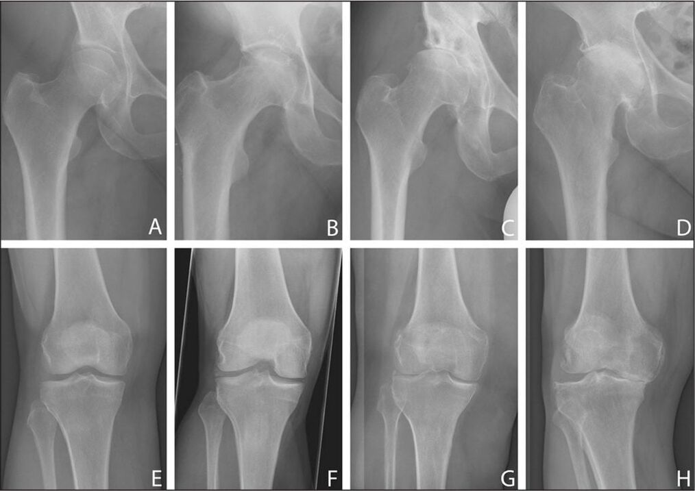
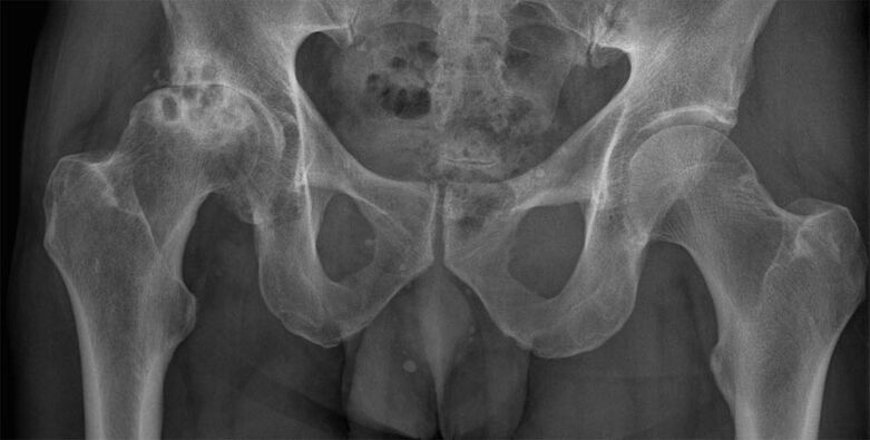
The stage of coxarthrosis is determined by X-ray examination. The resulting images clearly show destructive changes in the hip joint. This is a narrowing of the joint space, deformation of bone surfaces and formation of osteophytes.
computed tomography
Patients are prescribed a CT scan to determine the degree of flattening and deformation of the hyaline cartilage. The results of the study also make it possible to evaluate the state of the ligamentous-tendinous apparatus, nerve trunks, muscles and small and large blood vessels.
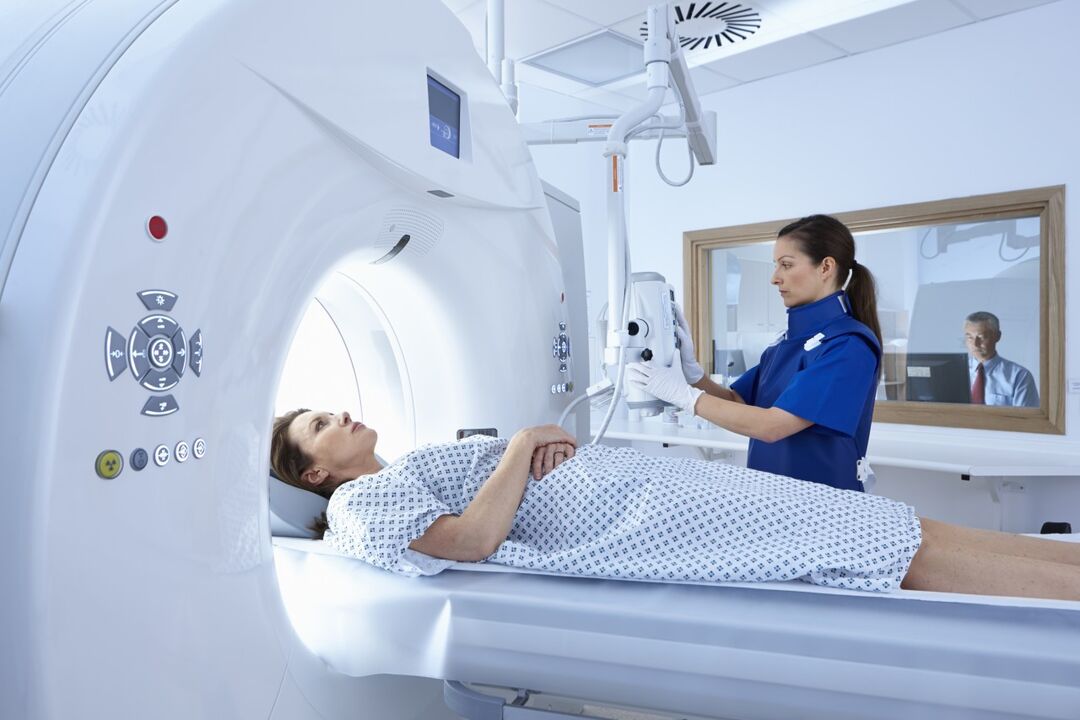
MRI image
Magnetic resonance imaging is one of the most informative studies in the diagnosis of coxarthrosis. To identify circulatory disorders in the affected joint area, contrast is performed. A routine study is prescribed to determine the degree of ligament damage and deformation of the femoral head, and to detect areas of fibrous degeneration of the joint capsule.
leg length measurement
Before the measurement, the doctor asks the patient to stand up and stretch his legs as much as possible. To obtain the most reliable data, the orthopedist uses two bone reference points. Upper: the anterior shaft of the pelvic bone, located on the anterior lateral surface of the abdomen at the outer edge of the inguinal ligament. The second reference point is any bony structure of the knee, ankle or heel. Measuring leg length may not be informative if coxarthrosis affects two hip joints at once.
laboratory research
Clinical blood and urine tests are performed to evaluate the patient's general health status. And the results of biochemical studies often make it possible to detect pathologies that provoked the development of coxarthrosis. Gouty arthritis is indicated by elevated levels of uric acid and its salts. An increase in the erythrocyte sedimentation rate and an increase in the number of leukocytes indicate the appearance of an inflammatory process (bursitis, arthritis, synovitis). To exclude rheumatoid arthritis, rheumatoid factor, C-reactive protein, and antinuclear antibodies are determined.

hip puncture
Synovial fluid is collected through puncture to study its composition and detect changes in its consistency. If an infectious inflammatory process is suspected, additional biochemical examination of a biological sample is indicated.
Treatment options
When determining treatment tactics, the orthopedist takes into account the severity of coxarthrosis, the form of its course, the causes of its development and the severity of symptoms. Patients are often recommended to wear rigid rib bandages and orthoses from the first days of treatment. Wearing orthotics helps slow cartilage degradation and bone deformation.
Medicines
Drugs from various clinical and pharmacological groups are used in the treatment of deforming osteoarthritis. These are non-steroidal anti-inflammatory drugs (NSAIDs), muscle relaxants, glucocorticosteroids, chondroprotectors, ointments and gels with a warming effect.
Blocking
To relieve acute pain that cannot be eliminated with NSAIDs, intra-articular or peri-articular pharmacological blocks are prescribed. To carry them out, hormonal agents are used. The analgesic effect of glucocorticosteroids is enhanced by their combination with anesthetics.
Injections
Intramuscular injection of NSAID solutions allows you to eliminate severe pain in the hip joint. To relax skeletal muscles, a drug is usually used that, in addition to a muscle relaxant, includes an anesthetic. In the form of injections, therapeutic regimens include vitamin B, drugs to improve blood circulation and chondroprotectors.
Diet therapy
Overweight patients are recommended to lose weight to stop the spread of pathology to healthy joint structures. The calorie content of the daily menu should be limited to 2000 kilocalories, excluding foods rich in fats and simple carbohydrates. Nutritionists recommend that all patients with coxarthrosis follow proper nutrition. The diet should contain fresh vegetables, fruits, berries, cereal porridge, fatty sea fish and dairy products. Following a therapeutic diet stimulates the strengthening of the immune system and improves general health.
Exercise and massage therapy.
Classic, acupressure and vacuum massages are used in the treatment of coxarthrosis. After several sessions, blood circulation in the hip joint improves and nutrient reserves are replenished. Carrying out massage procedures stimulates the strengthening of the ligamentous-tendinous apparatus and the restoration of soft tissues damaged by the displacement of osteophytes.
Regular exercise therapy is one of the most effective ways to treat osteoarthritis. A physiotherapist develops a set of exercises individually for the patient, taking into account her physical condition.
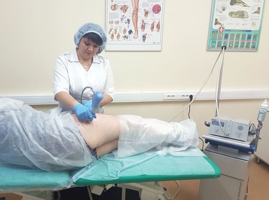
Physiotherapy
Patients with coxarthrosis are prescribed up to 10 sessions of magnetic therapy, laser therapy, UHF therapy, UV irradiation and shock wave therapy. The therapeutic effect of the procedures is due to the improvement of blood circulation, acceleration of metabolism and regeneration processes. To relieve acute pain, electrophoresis or ultraphonophoresis is performed with glucocorticosteroids, anesthetics and vitamin B. Applications with ozokerite or paraffin help eliminate discomfort.
Surgical intervention
If conservative treatment is ineffective, pain that cannot be eliminated with medication, or constant progression of coxarthrosis, patients are recommended to undergo surgical intervention. The operation is performed immediately in case of pathology of the third degree of severity, since it is impossible to eliminate the resulting destructive changes in cartilage and bones by taking medications or exercise therapy.
Arthroplasty
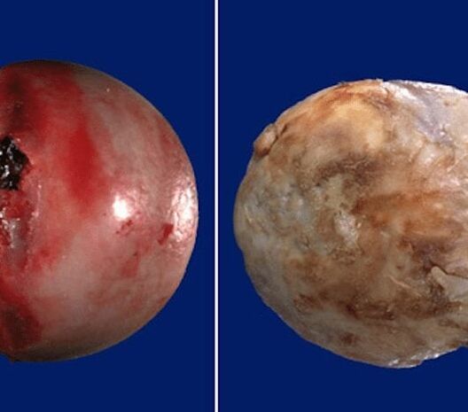
The operation is performed under general anesthesia. The head of the femur is removed from the acetabulum. Visible destructive changes in the tissue are corrected: bone growths are removed, joint surfaces are leveled, and tissue that has undergone necrosis is removed. During surgery, cavities are formed and filled with ceramic implants.
endoprostheses
Hip replacement with an implant is performed under general anesthesia. To prevent the development of an infectious process, antibiotic treatment is prescribed. After 10 days, the sutures are removed and the patient is discharged from the medical center. At the rehabilitation stage, patients are shown physiotherapy procedures and massage and exercise therapy.
Possible consequences
In the final stage of the pathology, flexion and adduction contractures develop. The patient's leg is constantly bent, so he uses a cane or crutches to move. After complete fusion of the joint space, immobility occurs, the patient cannot perform household chores and becomes disabled. Coxarthrosis is often complicated by aseptic necrosis of the femoral head, arthrosis of the knee joints, and arthritis.
Prevention and prognosis
Only grade 1 coxarthrosis responds well to conservative treatment. In other cases, endoprostheses make it possible to completely restore the functional activity of the hip joint. After installation of the stent, the patient quickly returns to an active lifestyle.
To prevent the disease, orthopedists recommend quitting smoking, abusing alcoholic beverages, doing daily physiotherapy and gymnastics, and, if necessary, losing weight.